Electroneuromyography
If you have any questions about the electrophysiological study, you can ask the doctor before the study begins. Below is an overview of the electrophysiological methods.
The electrophysiological study (electroneuromyography, ENMG) is primarily needed to determine the level of the lesion in the peripheral neuromuscular apparatus – involvement of a peripheral nerve, spinal cord motor neurons, the neuromuscular junction, or muscle fiber pathology. In polyneuropathy, the method allows differentiation between involvement of the myelin sheath – demyelinating neuropathy – or the axon – axonal neuropathy – and also confirms generalized or multifocal involvement of peripheral nerves in the pathological process. This group of techniques allows differentiation of motor root involvement in the pathological process.
The method is associated with unpleasant subjective sensations during electrical stimulation, but is generally well tolerated by most subjects. Its only absolute contraindication is the presence of purulent-inflammatory changes at the site of stimulation and electrode placement; it is not contraindicated in patients with concomitant cardiovascular disease or an implanted pacemaker.
The set of electrophysiological techniques includes stimulation techniques (measurement of nerve conduction velocity (NCV) along motor and sensory fibers, F-wave study (testing NCV at the most proximal segment of the nerve), detection of conduction block (CB), and, in rare cases, evaluation of the H-reflex), as well as needle electromyography – assessment of motor unit action potential parameters and spontaneous and insertional muscle fiber activity. A standard decrement test and jitter analysis are used to evaluate the state of the neuromuscular junction. Needle electromyography involves inserting a concentric needle electrode into the muscle and is accompanied by unpleasant sensations from the needle insertion, but no electrical stimulation is applied. The risk of complications from this procedure is comparable to that of a standard venipuncture.
Stimulation EMG. During stimulation EMG of motor fibers, the recording electrodes are placed over the corresponding muscle (the "belly-tendon" principle). The corresponding nerve is stimulated at different levels, and the summed electrical activity of the muscle under study is recorded and displayed by the electromyograph on a voltage-time scale as an M-wave. The distance between the two stimulation points and the difference in M-wave latency make it possible to determine the nerve conduction velocity (NCV) between the two stimulation points. The interval from the moment of the stimulus to the appearance of the first motor response is called the terminal (distal) latency. This parameter is not equivalent to the NCV in the distal nerve segment, since it does not account for synaptic delay time. The evoked motor response begins with a rising phase, repolarization, and hyperpolarization (figure 1), reflecting the asynchronous activation of muscle fibers during supramaximal nerve stimulation at varying distances from the recording skin electrodes.
 Waveform of the motor and sensory response during stimulation of the median nerve at different points along the arm.
During peripheral nerve stimulation, in addition to orthodromic propagation of excitation, there is also antidromic propagation to anterior horn neurons via interneurons. Activation of motor neurons leads to the appearance of a late response (F-wave). This response provides approximate information about NCV in proximal nerve segments that are inaccessible to direct stimulation (figure 2). The electrophysiological equivalent of the tendon stretch reflex is the H-reflex, which can be recorded by repeated stimulation of the common tibial nerve at 20-second intervals with recording of the late response from the soleus muscle.
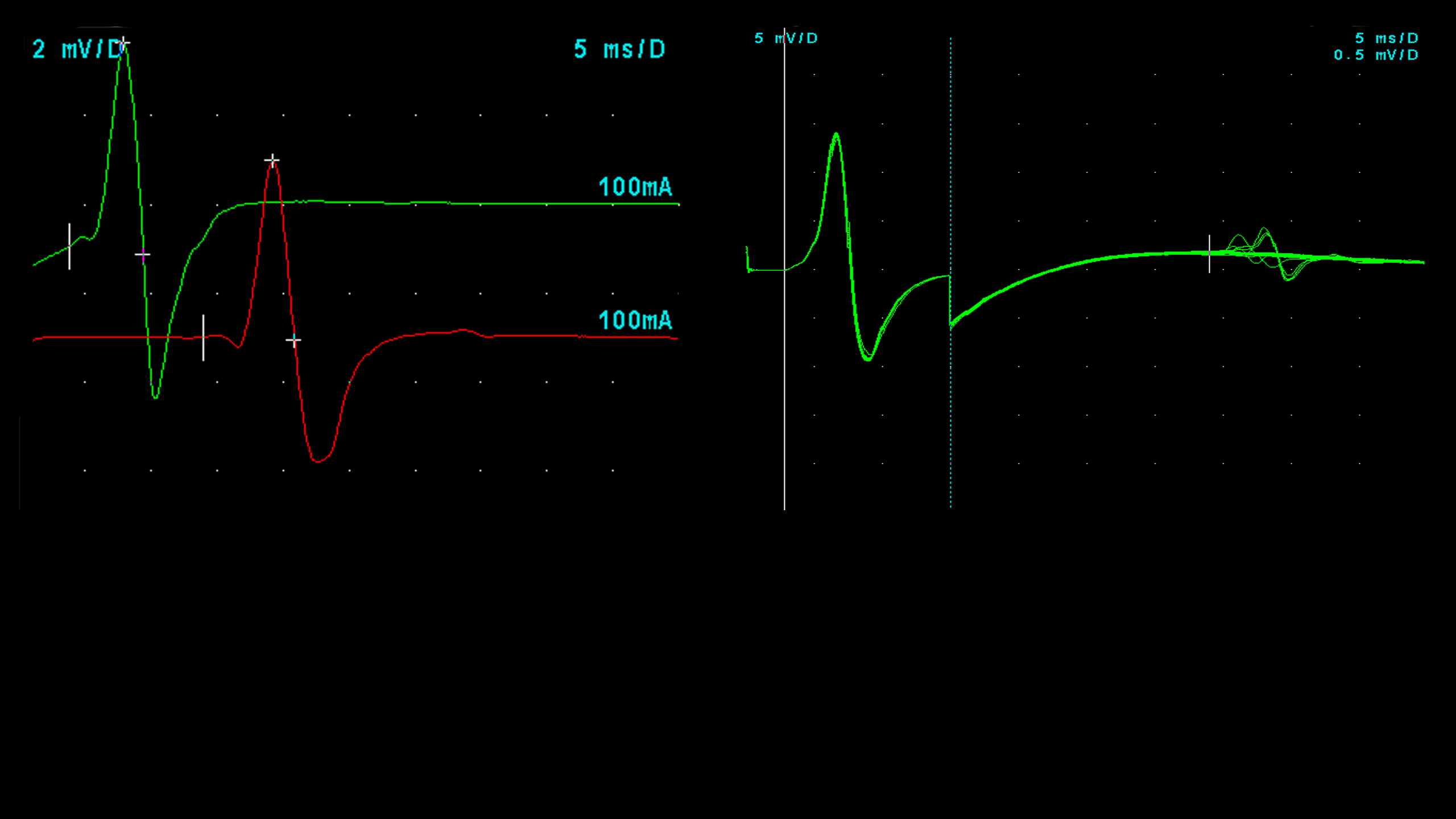 A. Evoked motor response during stimulation of the tibial nerve at the ankle and popliteal fossa levels. Recording from the Abductor Hallucis muscle.
B. F-wave during stimulation of the tibial nerve (superimposed traces)
A. Evoked motor response during stimulation of the tibial nerve at the ankle and popliteal fossa levels. Recording from the Abductor Hallucis muscle.
B. F-wave during stimulation of the tibial nerve (superimposed traces)
The nerve action potential (NAP), also called the evoked sensory potential, can be recorded by rhythmic low-amplitude stimulation of a peripheral nerve with recording from the skin surface (cutaneous receptors) in the corresponding innervation zone (antidromic technique), or, conversely, by stimulating skin surface receptors with recording from the nerve (orthodromic technique). The amplitude of the NAP and its NCV are assessed.
Needle EMG. To assess the electrical activity of a muscle, a concentric needle electrode is inserted directly into the muscle. Since the electrode samples only a small area of the muscle, it is moved to several points to increase the objectivity of the assessment (figure 3). This technique has a single absolute contraindication – the presence of purulent-inflammatory changes at the examination site. It is performed with caution (under INR monitoring) in patients on anticoagulant therapy, excluding paraspinal muscles from the study whenever possible. The risk of complications with this technique in other conditions, including in patients with a pacemaker, is comparable to that of a standard venipuncture.
Needle myography involves three stages. In the first stage, spontaneous and (insertional) activity of muscle fibers is assessed with the muscle fully relaxed.
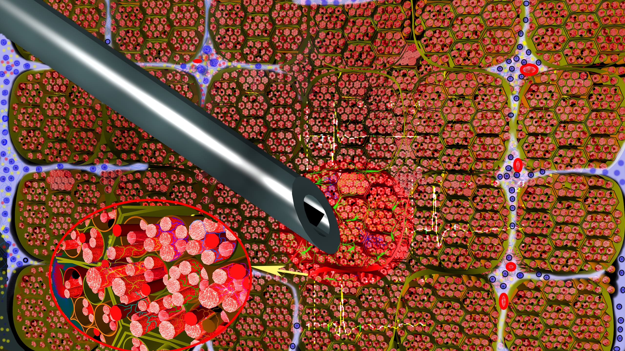 Recording of a motor unit action potential using a concentric needle electrode.
 Needle electrode in the muscle during ultrasound examination.
1. Bundles of muscle fibers
2. Axon terminals
3. Needle electrode
4. Epimysium
5. Perimysium
6. Recording area of the concentric electrode
7. Arterioles
Insertional activity ("needle insertion potential") occurs within 0.5–1 ms as a brief electrical burst caused by mechanical irritation of muscle fibers by the needle electrode. It may be increased, decreased, or include wave phenomena – the end-plate potential (end-plate noise) – an electrical potential of the muscle fiber membrane resulting from irritation of excitation from axon terminals during their mechanical stimulation by the needle electrode, along with high-frequency discharges. Experiments in rats have shown increased end-plate noise amplitude together with recording of fibrillation potentials and positive sharp waves at a myofascial trigger point of a muscle. These findings have provided grounds for considering end-plate dysfunction as the main cause of myofascial trigger point formation.
Under normal conditions, outside the end-plate zone, a resting muscle has no electrical activity. Therefore, any activity in a relaxed muscle is considered pathological. Based on electrophysiological characteristics, several types of spontaneous activity are distinguished – fibrillation potentials (FP), positive sharp waves (PSW), fasciculation potentials (FasP), complex repetitive discharges (CRD), myotonic discharges, neuromyotonic discharges, myokymia, and cramps. All of these phenomena are non-specific and may develop in both neurogenic and myopathic processes.
Fibrillation potentials (FP) have a biphasic or triphasic shape with an initial positive or negative deflection and a duration of 1 to 5 ms. They arise in muscle fibers that have lost their innervation in neurogenic conditions, or as a result of fiber splitting and separation from the end-plate zone in muscle pathology.
Positive sharp waves (PSW) have a biphasic shape with an initial sharp positive phase followed by a prolonged negative phase lasting 10 to 30 ms. Their causes are similar to those of FP.
Spontaneous depolarization of a muscle fiber membrane, with subsequent spread of excitation to neighboring muscle fibers, leads to the development of a complex repetitive discharge (CRD). Subsequently, a varying number of neighboring muscle fibers may be sequentially depolarized until the "circuit" is complete, after which the original muscle fiber discharges again. Each spike in this discharge belongs to a separate muscle fiber, which in turn may be part of different motor units but located close to one another. This electrophysiological phenomenon has a regular pattern with an abrupt onset and cessation and a frequency of 3 to 40 Hz, with a characteristic sound (like the sound of a boat motor).
Myotonic discharges are action potentials of individual muscle fiber membranes with an exponentially changing frequency (40–100 Hz) and amplitude, producing a characteristic sound (like a "dive bomber"). Although slow myotonic phenomena resemble FP, the distinguishing feature is the rapid rate of change in frequency and amplitude. These phenomena are characteristic of myotonic dystrophy, myotonia, and paramyotonia, and may also occur in other muscle diseases without myotonia (hyperkalemic paralysis, polymyositis, etc.), rarely in axonal neuropathies, but they are never the predominant form of spontaneous activity in such cases.
Fasciculation potentials (FasP) are spontaneous discharges of single motor units that can be generated anywhere along the lower motor neuron. Their electrophysiological characteristics are identical to the recording of a single motor unit potential with an irregular firing rate. They usually occur in neurogenic disorders (motor neuron disease, axonal neuropathies).
Myokymic discharges are a group of spontaneously occurring, repeating bursts of motor unit action potentials at a frequency of 40–60 Hz, followed by a brief period of electrical silence (0.1–10 sec), with the same sequence repeating. They have a regular, semi-rhythmic pattern and sound (resembling "marching soldiers"). They are most often seen in radiation nerve damage and chronic compressive neuropathies.
Neuromyotonic discharges are bursts of motor unit action potentials arising in motor axons and firing at high frequencies (100 to 300 Hz). They repeat either continuously or as repeating bursts of decreasing amplitude. They are not affected by voluntary activity. They usually occur due to a defect in voltage-gated potassium channels and are typically seen in disorders of increased nerve excitability (Isaacs syndrome, Morvan syndrome).
Cramp potentials are involuntary, repetitive, high-frequency recruitment of motor unit action potentials over a large area of muscle. They usually build up rapidly with the addition of new potentials and end abruptly. They can be observed in healthy people during excessive muscle activation, as well as in any chronic neurogenic disorder, electrolyte disturbances, or increased peripheral nerve excitability.
Voluntary muscle activity is mediated by the function of the lower motor neuron and the muscle fibers it innervates. This complex constitutes the concept of the motor unit action potential (MUAP). During voluntary muscle contraction, MUAPs are recorded in a semi-rhythmic pattern at a frequency dependent on the degree of voluntary muscle activation. A concentric needle electrode can only detect MUAPs located near its recording surface (generally within 0.5 mm). Recruitment of MUAPs is the activation of additional motor units as the firing rate of the initial (active) MUAP increases. The summed electrical potential of all available motor units recorded by the concentric electrode per unit time at the muscle point under study, displayed on a voltage-time scale, is called the interference pattern.
The method of isolating and analyzing voluntarily recruited, repeating MUAPs with evaluation of their amplitude, duration, and number of phases is called quantitative EMG. Because of the large number and variety of MUAP parameters, this method requires multiple measurements and statistical description of results obtained from different muscle sites. Advances in digital technology have made it possible to perform computerized algorithmic template matching, known as quantitative EMG decomposition (QEMG), which has significantly improved the reliability and reproducibility of muscle electrical activity parameters. Each MUAP is characterized by rise time (the time of rapid positive or negative deflection from the baseline), duration (the time from the first deviation from the baseline to its final return), amplitude (the maximum peak-to-peak amplitude of the main potential), number of phases (the number of times the potential crosses the baseline, plus one), and stability (any change in MUAP configuration in the absence of electrode movement).
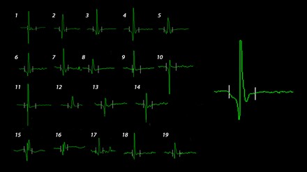 19 voluntarily recruited MUAPs from different points of the tibialis anterior muscle of a healthy subject, with the analyzed parameters – amplitude, duration, and phase (right).
In neuromuscular pathology, MUAP parameters change, which, combined with the recording of spontaneous activity, allows the examiner to determine the level of the lesion. Loss of muscle fibers across a wide range of muscle pathologies leads to a decrease in MUAP duration, an increase in the number of phases, and, in some cases, a decrease in amplitude. At the same time, compensatory hypertrophy of the muscle fiber can produce individual high-amplitude potentials. Spontaneous activity in these cases arises on a damaged or unstable muscle fiber membrane during its splitting or breakdown. Loss of a significant number of axons leads to a decrease in the number of active motor units and a decrease in interference pattern frequency, followed by compensatory reinnervation of muscle fibers deprived of nerve control through collateral sprouting. This leads to increased amplitude and duration, subsequently to polyphasia and loss of motor unit stability. A muscle fiber that has lost its connection to the axon generates an electrical signal on the membrane surface, recorded as spontaneous activity. In this case, the electrophysiological characteristics of spontaneous activity do not differ between primary muscle and denervation processes. Questions about the mechanism of MUAP parameter changes in different types of lesions are beyond the scope of this monograph and are discussed in detail in the works of Buchthal F and Stålberg E.
Thus, neurophysiological assessment methods are essential in evaluating patients with various levels of peripheral neuromuscular apparatus involvement. The method requires the examiner to control various technical factors, possess data-collection skills, and understand signal changes that may be caused by a wide range of causes. Our center performs the full range of electrophysiological techniques at a high level, and we are ready to offer an individual approach to your problem and ensure compliance with all generally accepted standards.
Author of the text: MD, PhD Dmitry S. Druzhinin