Confocal Microscopy of Nerves
The potential of confocal microscopy (CM) for assessing C-fibers in peripheral neuropathy is increasingly being investigated. It is a non-invasive procedure for evaluating the terminal fibers of the ophthalmic branch of the trigeminal nerve — the corneal nerves — which are part of the peripheral nervous system. At a distance of approximately 1 millimeter from the limbus, these fibers lose their myelin sheath to ensure corneal transparency and terminate in the epithelium of the superficial cells as small invaginations. The main parameters analyzed are nerve fiber density and length, as well as counting the number of dendritic and non-dendritic Langerhans cells. A normative database has been established in a population of healthy individuals.
Currently, three CM modes are used: laser scanning, slit-scanning, and tandem scanning CM. These modes differ in light source, magnification, contrast, and resolution. The use of the method has been described in diabetic neuropathy, idiopathic small fiber neuropathy, Fabry disease, and a number of conditions causing small fiber damage, including hereditary sensory and autonomic neuropathy (HSAN), dysimmune neuropathy, Crohn's disease, chemotherapy-induced neuropathy, and trigeminal neuralgia.
Of particular interest is research assessing the degree of damage to thin unmyelinated fibers based on confocal microscopy findings in patients with hereditary motor and sensory neuropathy type 1A. Changes in the density of corneal nerve distribution have also been shown in ocular diseases (dry eye syndrome, keratoconus, contact lens wear, neurotrophic keratopathy, recovery of sensitivity after corneal surgery, etc.). Changes in corneal nerve density have also been noted in patients with chronic migraine and ischemic brain injury.
Figure. Confocal microscopy method. Demonstration of the confocal microscopy method for thin C-fibers.
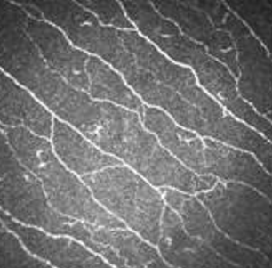
Thus, confocal microscopy of the corneal nerves is a non-invasive, accessible method that objectively assesses the degree of involvement of thin unmyelinated C-fibers, opening up the possibility of early subclinical diagnosis of neuropathies of various origins. The search for nosologically specific signs has not yet been successful, despite the technological advancement of the method.
Text author: MD, DSc Дружинин Д.С.